Electron Energy-Loss Spectroscopy: The “Swiss Army Knife” of Electron Microscopy
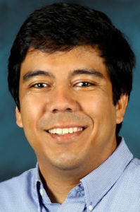
Prof. Juan Carlos Idrobo, Oak Ridge National Laboratory
Abstract

Electron energy-loss spectroscopy (EELS) in scanning transmission electron microscopy (STEM) is arguably one of the most versatile spectroscopy techniques to study materials at (sub)nanometer length scales. For many decades, EELS has been used to measure sample thickness, obtain elemental chemical compositional maps and extract the bonding information of materials with unprecedented spatial resolution [1]. Improvements in the understanding of the scattering process involved in EELS have also resulted in the ability of EELS to measure and quantify ferromagnetic phases in materials, as it is done with X-ray circular dichroism in synchrotrons, but with the phase of the electrons playing the role of polarization of light [2]. The recent development of a new generation of monochromators [3] and spectrometers [4] has made EELS even more versatile. Phonon mapping with atomic resolution [5,6], primary temperature measurements at the nanoscale (without requiring any previous knowledge of the sample) [7], and the detection of isotopes in water [8] and amino acids [9] are now some of the new “tools” in this Swiss Army knife of spectroscopy that is EELS. Here, we will review the technical details of the aforementioned tools. We will discuss potentially relevant new “blades” that could be added to the EELS toolkit, to accelerate the discovery of new physical phenomena and sharpen our understanding of how mater behaves [10, 11].
[1] R. Egerton Electron Energy-Loss Spectroscopy in the Electron Microscope, 3rd edition, (Springer, 2011); [2] P. Schattschneider, et al., Nature 441 (2006) p. 486; [3] O. L. Krivanek, et al., Phil. Trans. R. Soc. A 367 (2009), p. 3683; [4] T.C. Lovejoy et al., Micros. & Microanal. 24 (2018) p. 446; [5] K. Venkatraman et al., Nat. Physics 15 (2019) p. 1237; [6] F. S. Hage et al., Science 367 (2020), p. 1124; [7] J. C. Idrobo, et al, Phys. Rev. Lett. 120 (2016), p. 095901; [8] J.R. Jokisaari, et al., Adv. Mater. 30 (2018), p. 1802702; [9] J.A Hachtel, et al., Science 363 (2019), p. 525; [10] J.C. Idrobo, Nat. Rev. Mater. 6 (2021), p. 100; [11] This research was supported by the Center for Nanophase Materials Sciences, which is a Department of Energy Office of Science User Facility. This research was conducted, in part, using instrumentation within ORNL’s Materials Characterization Core provided by UT-Battelle, LLC under Contract No. DE-AC05-00OR22725 with the U.S. Department of Energy.
Biosketch

Juan Carlos Idrobo is a Senior Staff Scientist at Oak Ridge National Laboratory and Group Leader of the Scanning Transmission Electron Microscopy Group at the Center for Nanophase Materials Sciences. His research consists in applying analytical techniques in electron spectroscopy within monochromated and aberration-corrected scanning transmission electron microscopy to study the structure, electronic, magnetic, thermal, optical and topological properties of materials. He has published over 170 papers in peer review journals with over 18,000 citations. In 2018 Idrobo was recognized by Clarivate Analytics to be among the top 1% of researchers being cited in Cross-Field in Web of Science, between 2006 and 2016. Idrobo holds Physics degrees from Universidad de Los Andes in Colombia (B.Sc., 2000), University of Illinois at Chicago (Master, 2003) and University of California Davis (Ph.D., 2004).
